Introduction
Airborne microorganisms are microorganisms that can be transported via the air currents and dust particles. It can cause the contamination easily and must be aware by all microbiologist. The procedure for the assay must be done carefully and the precaution should be taken.
Airborne particles are a major cause of respiratory ailments of humans, causing allergies, asthma, and pathogenic infections of the respiratory tract. Airborne fungal spores are also important agents of plant disease, and the means for dissemination of many common saprotrophic (saprophytic) fungi.
One of the type of airborne microorganisms is resident microorganisms. Resident microorganisms are your own body's defence mechanism, also called normal flora. They are bacteria that live all over your skin and fight off other pathogenic microorganisms from invading you so to say. However, the opportunity pathogens will infect our body and cause a disease occurs when we are injure or when our immune system are weakened.
Another type of airborne microorganisms is transient microorganisms. These microorganisms include food-borne microorganisms and even soil-borne microorganisms that make their way into the human digestive tract and, depending upon the characteristics of the specific organism involved, either subtly or dramatically influence the overall health of the human system. Transient microorganisms are different from resident microorganisms in that they do not take up permanent residence in the gastrointestinal tract. Instead, they establish small colonies for brief periods of time before dying off or being flushed from the intestinal system via normal digestive processes, and/or by peristaltic bowel action.
At the upper respiratory nasal cavity, there are either normal residents or transients. However, normal microflora have a large number of non-pathogenic or opportunistic pathogens on our skin. Since most of the transient microorganisms are pathogenic, and it can comes in our body via respiration and when we eating. So, we need to clean our hand properly before we eat and during food preparation to minimise the opportunity of eating the pathogenic microorganisms.
Objective
To determine the microorganisms in the air and from healthy humans.
Materials and reagents
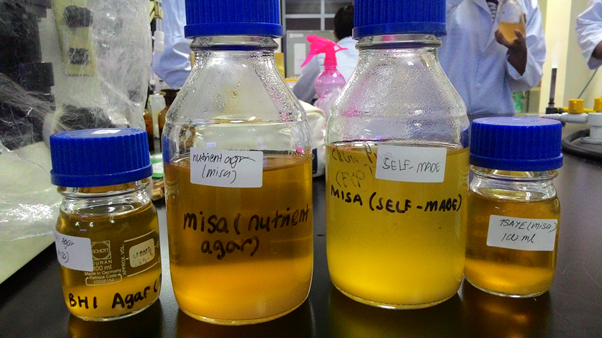
Molten nutrient agar
Sterile water
Sterile petri dishes
Sterile clinical swab
Pipette and tips
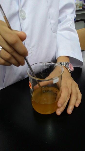
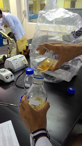
Procedure
(REFER LAB MANUAL)
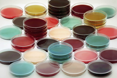
Results
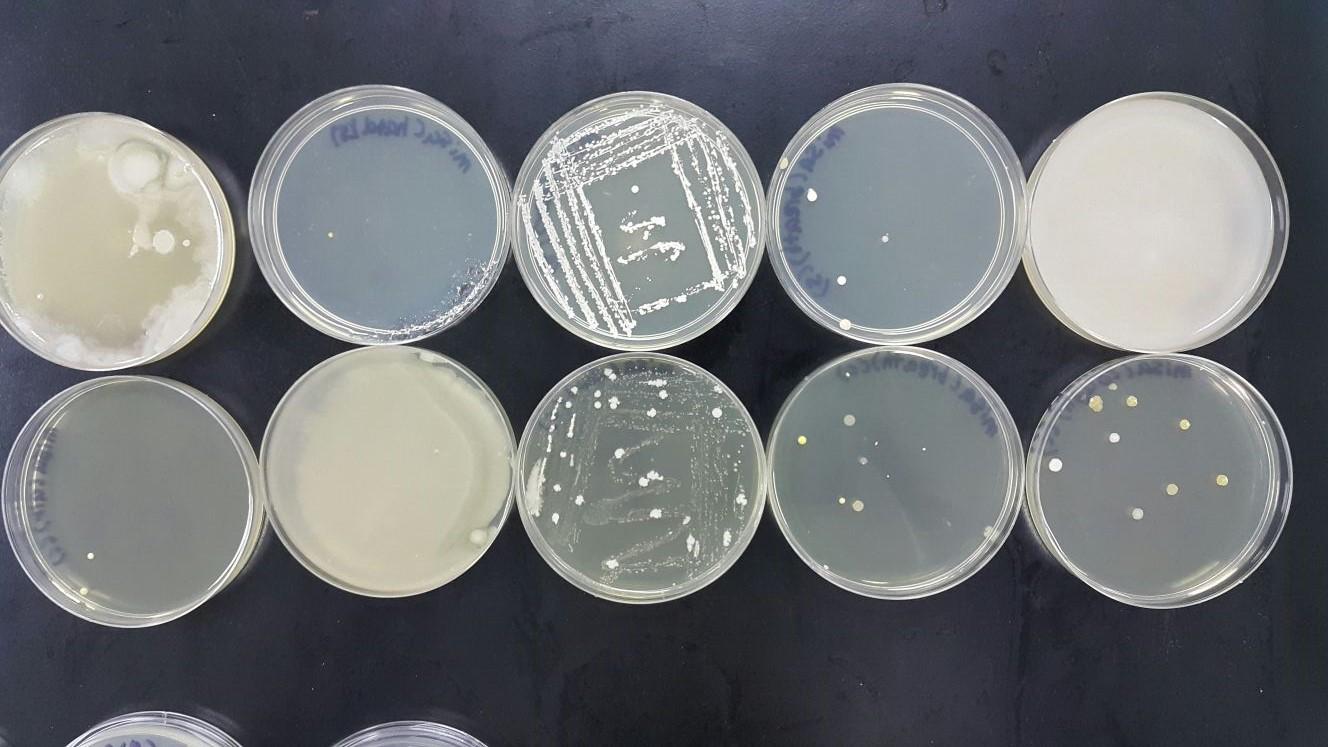
Colonies from air
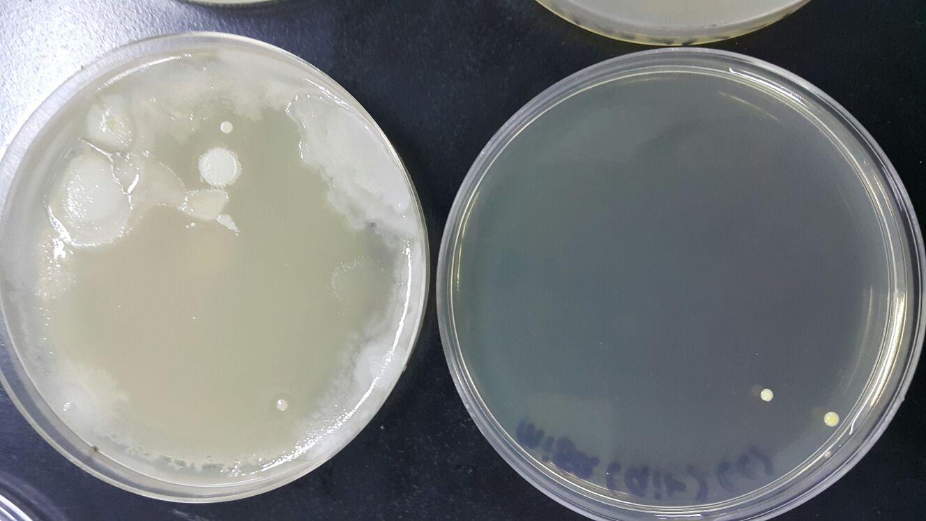
Colonies from hands
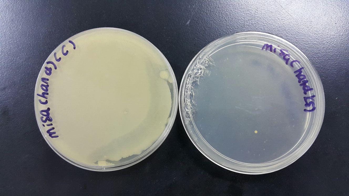
Colonies from ears

Colonies from normal breathing

Colonies from violent coughing
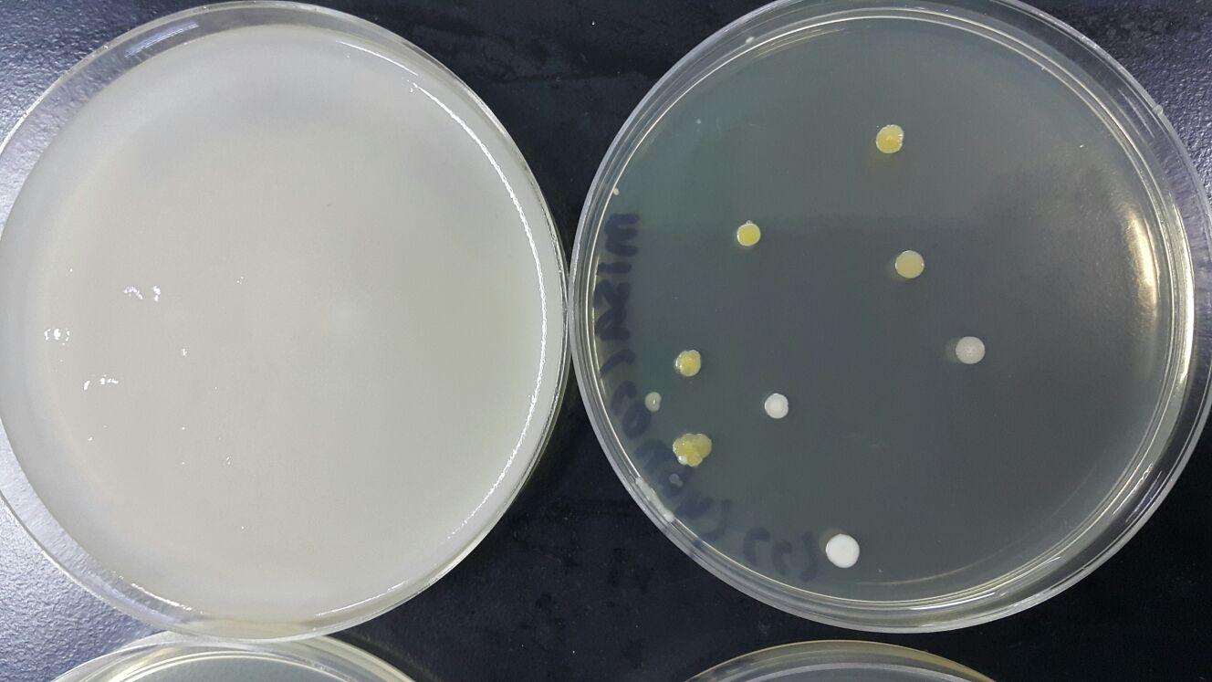
All petri dish after incubate at 37°C for 48 hours
Colonies From
|
Self-made Nutrient Agar
|
Commercial Nutrient Agar
|
Air
|
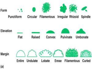
Circular, flat, shiny smooth, entire
|
Circular, flat, shiny smooth, entire
|
Normal Breathing
|
Irregular, flat, umbonate , smooth, undulate
|
Irregular, flat, shiny smooth, undulate, filiform, rough, dull
|
Ear
|
Irregular, filiform, flat, shiny smooth, entire
|
Irregular, filiform, flat, shiny smooth, entire
|
Violent Cough
|
Irregular, round, flat, shiny smooth, entire, raised
|
Circular, cruteriform
|
Hands
|
Filamentous, circular, entire, flat, shiny smooth
|
Filamentous, circular, entire, flat, shiny smooth
|
(Figure 1: Morphology of the colonies)
Discussion
Bacteria populations grow extremely fast under the desired nutrients and environmental conditions. Different types of bacteria will produce colonies that are distinctive in appearance. Colony morphology is a method that scientists use to describe the characteristics of an individual colony of bacteria growing on agar in a Petri dish. It can be used to help to identify them .Each distinct colony represents an individual bacterial cell or group that has divided repeatedly. Being kept in one place, the resulting cells have accumulated to form a visible patch.
- We use the following guide to determine the morphology of the colonies:-
- The atmosphere is not a very welcoming environment for many microorganisms. The joint effects of desiccation and sunlight cause many microbial cells to die rapidly when suspended in air. This is especially true of Gram-negative bacteria, including food borne pathogens like E. coli and Salmonella. Nevertheless, some Gram-positive bacteria and fungal spores can survive for long periods in the atmosphere and can be widely dispersed by air currents. The typical microflora of the air is usually made up of pigmented Gram-positive bacteria and bacterial and fungal spores, which are resistant to the drying effects of the air and to radiation. Unfortunately, it can include some pathogenic bacteria, such as Staphylococcus aureus and Bacillus cereus, and common food spoilage fungi.
- Our mouth contains a lot of pathogenic and non-pathogenic microorganisms. The pathogenic bacteria will cause some microbial diseases of the respiratory system which may occur in the upper or lower regions. Some examples of these non-pathogenic bacteria are Streptococcus, Neisseria, Haemophilus, and Micrococcus. Whereas the pathogenic bacteria might cause strep throat, scarlet fever, diptheria.
- A cough is a sudden and often repetitively occurring reflex which helps to clear the large breathing passages from secretions, irritants, foreign particles and microbes. Actually the microbes during breathing and coughing are quite similar because there are originated from the same place, mouth and nose cavity in human beings.
- The hands and fingernails are often affected by fungal and yeast infections. Some of the species are Aspergillus, Acremonium, Epidermophyton,and Trichophyton. There are also some bacteria that exist on our hands. These are the few common examples, Serratia, Aeromonas, Klebsiella
Conclusions
Bacteria are everywhere and can spread from surface to surface, person to person, food to food, and person to food. Harmful bacteria can be controlled by practicing the 4 Cs of food safety. To prevent the spread of harmful bacteria, proper cleaning of both hands and surfaces is especially important. The good thing is that not all bacteria are harmful; most bacteria are beneficial to us.
When designing experiments, you should always use safe techniques when working with bacteria. Also, it's important to have a control plate. In this experiment, you also learned that different strains of bacteria can be identified through colony morphology.
References
http://www.scientificpsychic.com/health/hygiene.html
http://www.cliffsnotes.com/study_guide/Bacterial-Diseases-of-
http://microbewiki.kenyon.edu/index.php/Human_Hands_and_Fingernails
http://www.wisegeek.com/what-is-bacterial-contamination.htm
http://www.cliffsnotes.com/study_guide/Bacterial-Diseases-of-
http://microbewiki.kenyon.edu/index.php/Human_Hands_and_Fingernails
http://www.wisegeek.com/what-is-bacterial-contamination.htm
http://allnurses.com/nursing-student-assistance/whats-the-difference-423438.html
http://www.relfe.com/lactobacillus.html
http://www.relfe.com/lactobacillus.html