2.1 Ocular Micrometer
Introduction
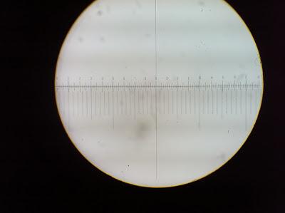
Ocular micrometer is used to measure the size of magnified objects. It is a glass disk that has a ruled scale which fits in a microscope eyepiece. The physical length of the marks on the scale depends on the degree of magnification. Micrometer is a scale or ruler which on a flat circle of glass. Before we use the ocular micrometer, we need to insert it into one of the microscope eyepiece. The stage micrometer is necessary used to calibrate it for each of the objectives before using the ocular micrometer. A direct measurements of an observed object can be made during the calibration of the stage micrometer with the ocular micrometer. After that, the line superimposed certain distance markers on the microscope field. The stage micrometer is a microscope slide which has a finely divided scale marked on the surface and is used for calibration of optical system with eyepiece graticule patterns. By determining how many units of the ocular micrometer superimpose a certain distance on the stage micrometer, we can calculate the distance between each ocular division measures on the microscopic field. This can help us calculate the object more accurate and precise. After we determine the units, we can take out the stage micrometer and put the specimen slide on the stage of microscope to determine its length and width.

(Figure 1: The scale of stage and ocular microscope)
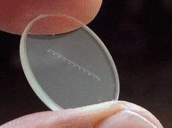
(Figure 2: The glass slide of ocular micrometer)
Objective
To learn how to measure and count the cells by using light microscope.
To learn the technique in calibration and calculation of the ocular micrometer.
Materials and Reagent
Light microscope fitted with an ocular micrometer
Stage micrometer
Stained preparation of yeasts and bacteria
Procedure
(Refer to lab manual)
Result
(400x magnification)
10 division on stage = 10x0.01mm
=0.1mm
10 division on stage = 40 division on ocular,
1 ocular = 0.1mm/40
= 0.0025 mm
Since 1 mm= 1000 μm
Thus,the size of yeast under 400x magnification= 0.0025 mm x 1000 = 2.5 μm
Discussion
An ocular micrometer is a glass disk with a ruled scale that fits into a microscope eyepiece. It is used to measure the size of objects. It use together with a stage micrometer for measurement with greater accuracy. The standard eyepiece reticle, when combined with a precision stage micrometer, provides a rapid, convenient, and accurate means of conducting measurements in the microscope.
We use a stage micrometer to calibrate the ocular micrometer. A stage micrometer is essentially a ruler that is mounted on a microscope slide that does have units (millimeters (mm) or micrometers (m)). When calibrating, we line up the stage micrometer with the ocular micrometer and count the number of divisions on the ocular micrometer per millimeter or micrometer on the staged micrometer. The number of divisions will change as the magnification changes.
2.2 Neubauer Chamber
Introduction
The Neubauer Chamber or hemocytometer is a specimen slide which its function is used to determine the cells’ concentration in a liquid sample. It was originally designed for counting of blood cells. Hemocytometer consists of a thick glass microscopic slide with a rectangular indentation that creates a chamber. The chamber is engraved with a laser-etched grid of perpendicular lines. The device is precisely crafted so that the area bounded by the lines is known, and the depth of the chamber is also known. Thus, it is possible to count the number of cells or particles in a specific volume of fluid, and thereby deter the concentration of cells in the fluid overall.


Materials and Reagents
Diluted Yeast culture
Neubauer and coverslip
Sterile Pastuer pipettes
Procedure
(Refer to lab manual)
Results
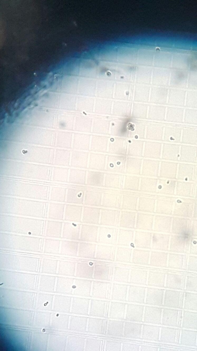
Image of yeast on hemacytometer (100x)
Average number of the cells per square box
= (2+2+2+4+5+5+4+2+5)/10
=4
Volume of one small box = 1mm x 1mm x 0.1mm
= 0.1mm^3
1mm = 0.001cm^3
0.1mm^3= 0.0001cm^3
1cm^3 = 1 ml
0.0001cm3 = 0.0001 ml
Volume of square = 250000 mL
Cell concentration= 4 cells x 250000 mL x (10^1dilution factor)
=1 x 10^7cells/mL
Discussions:
The hemocytometer consists of a thick glass microscope slide with a rectangular indentation that creates a chamber. This chamber is engraved with a laser-etched grid of perpendicular lines. The device is carefully crafted so that the area bounded by the lines is known, and the depth of the chamber is also known. Therefore it is possible to count the number of cells or particles in a specific volume of fluid, and thereby calculate the concentration of cells in the fluid overall.
The ruled area of the hemocytometer consists of several, large, 1 x 1 mm (1 mm2) squares. These are subdivided in 3 ways; 0.25 x 0.25 mm (0.0625 mm2), 0.25 x 0.20 mm (0.05 mm2) and 0.20 x 0.20 mm (0.04 mm2). The central, 0.20 x 0.20 mm marked, 1 x 1 mm square is further subdivided into 0.05 x 0.05 mm (0.0025 mm2) squares. The raised edges of the hemocytometer hold the coverslip 0.1 mm off the marked grid. This gives each square a defined volume. The cell-sized structures counted lie between the middle of the three lines on the top and right of the square and the inner of the three lines on the bottom and left of the square.
Conclusions:
The ocular micrometer allows us to measure the size of the specimen more precisely. By using the Neubauer chamber, we also learned the way to calculate the concentration of yeast cells of the sample we used.
References:
https://en.wikipedia.org/wiki/Ocular_micrometer
http://labreport102.blogspot.my/2014/11/lab-report-2-group-2.html
http://www.pyser-sgi.com/images/thumbnails/Graticules/Stage%20Micrometers%20web.pdf
http://elearning.usm.my/1516sem1/
academic.evergreen.edu/curricular/fcb/wk2calibration.doc
http://www.mecanusa.com/microscope/micrometer/micrometer1.htm
http://en.wikipedia.org/wiki/Hemocytometer
http://en.wikipedia.org/wiki/Hemocytometer
http://www.microbehunter.com/the-hemocytometer-counting-chamber/
No comments:
Post a Comment